
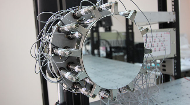
Researchers at Samara University have developed an innovative and robust digital image processing algorithm designed to significantly enhance vein visibility. The scientists envision this advancement as a critical tool to assist medical professionals in performing more accurate needle insertions and blood draws, particularly in cases where veins are not easily discernible to the naked eye. The detailed findings of this research have been published in the Journal of Biomedical Photonics & Engineering.
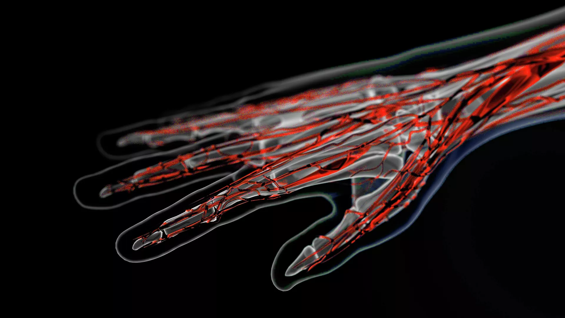
Image depicting veins on a hand.
Modern medical laboratory diagnostics frequently necessitate the insertion of a needle into a patient`s vein. However, blood vessels are often not visible to the unaided eye, making this medical procedure challenging for healthcare workers, for instance, when collecting blood samples.
To address this prevalent issue, scientists are actively advancing various optical methods, including near-infrared vein visualization, akin to night vision cameras. Nevertheless, these existing techniques present several limitations, such as shallow penetration depth and low image contrast. Furthermore, the digital image processing algorithms currently employed in these methods often suffer from instability and insufficient performance.
The researchers at Samara University have successfully created an advanced and stable digital image processing algorithm for superior vein visualization. Nikita Remizov, a postgraduate student at the Department of Laser and Biotechnical Systems at Samara University, explained that the algorithm is based on efficient operations derived from the Discrete Fourier Transform. This technique is widely used in signal digitization algorithms, with modifications applied in areas like audio compression (MP3) or image compression (JPEG).
“Image processing in the Fourier domain allows for effective and reliable enhancement of areas with sharp changes in pixel intensity. Regions located in the high-frequency domain of a two-dimensional spectrum correspond to these sharp transitions in the image. By utilizing the Fast Fourier Transform, we can employ relatively cost-effective computing modules and process images in real-time,” Remizov stated.
According to the researchers, this newly developed algorithm surpasses existing methods in its ability to accurately differentiate pixels corresponding to veins from those associated with surrounding bodily tissues.
“This new algorithm represents a crucial step in the development of a domestic vein visualization device. The aim is to create a device that is affordable to manufacture, even under sanctions, and is both effective and convenient for medical professionals when working with patients who have a high body mass index or other factors that complicate venipuncture,” Remizov emphasized.
Currently, the team is focused on optimizing the optical configuration of the forthcoming device. This, combined with the algorithmic image processing, is expected to enable the visualization of veins that are currently inaccessible using other methods, such as in cases of pronounced skin pigmentation.